| 1 |
Stereo Zoom Microscope |
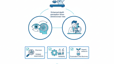
Uses two separate optical paths to provide a three-dimensional view of a specimen. The zoom objective changes magnification continuously without changing the working distance significantly. |
5×–80× |
4:1–8:1 |
50–200 mm |
Binocular or trinocular viewing; transmitted, reflected, or ring illumination. |
Assembly, dissection, education, electronics, biological specimens, and general inspection. |
Choose according to zoom range, optical resolution, stand stability, eyepiece field of view, and available accessories. |
| 2 |
Trinocular Stereo Zoom Microscope |
Works like a stereo zoom microscope but includes a dedicated camera port that allows simultaneous viewing through the eyepieces and image capture. |
7×–90× |
4:1–8:1 |
60–180 mm |
Binocular observation plus a camera port; coaxial, ring, gooseneck, or transmitted lighting. |
Quality control, documentation, training, research, and remote collaboration. |
Check camera-port compatibility, parfocal performance, sensor size, optical splitting ratio, and software support. |
| 3 |
Digital Zoom Microscope |
Uses a digital camera and a motorized or electronically controlled zoom lens to display magnified images on a monitor instead of relying mainly on eyepieces. |
10×–300× displayed |
4:1–10:1 optical zoom |
20–150 mm |
HD or 4K monitor output; adjustable LED ring, coaxial, or low-angle illumination. |
Production inspection, solder-joint checks, education, demonstrations, and inspection records. |
Separate optical magnification from digital enlargement; evaluate working distance, frame rate, latency, image calibration, and software functions. |
| 4 |
Video Inspection Zoom Microscope |
Combines a zoom optical system with a live video camera for continuous inspection on a screen, often with measurement, annotation, and image-capture functions. |
5×–200× displayed |
6:1–12:1 |
30–250 mm |
Live HDMI, USB, or network video; brightfield, darkfield, coaxial, and polarized illumination may be available. |
Incoming inspection, surface defects, printed circuit boards, machined components, and repair work. |
Prioritize image latency, measurement accuracy, field of view, lighting uniformity, and compatibility with existing inspection systems. |
| 5 |
Industrial Inspection Zoom Microscope |
Uses a robust zoom optical head mounted on a stable stand or articulated arm for repeatable viewing of manufactured parts and assemblies. |
6×–100× |
4:1–8:1 |
80–300 mm |
High-intensity LED, coaxial, darkfield, low-angle, or programmable lighting. |
Metal parts, plastics, connectors, welds, castings, coatings, and production-line quality control. |
Consider vibration resistance, stand reach, lighting control, inspection ergonomics, calibration, and serviceability. |
| 6 |
Digital Measurement Zoom Microscope |
Combines zoom optics, a camera, and calibrated software to measure dimensions, angles, radii, distances, and geometric features. |
10×–250× displayed |
4:1–10:1 |
30–200 mm |
High-resolution camera; telecentric or near-telecentric accessories may reduce perspective error. |
Dimensional inspection, tooling, micro-machined parts, stamped components, and production verification. |
Verify calibration traceability, pixel resolution, measurement uncertainty, stage accuracy, software features, and export formats. |
| 7 |
Polarizing Zoom Microscope |
Uses polarizer and analyzer filters to examine how transparent, translucent, or reflective materials interact with polarized light. |
5×–80× |
4:1–8:1 |
50–180 mm |
Cross-polarized or parallel-polarized illumination; rotating stage may be included. |
Minerals, crystals, plastics, fibers, glass stress, coatings, and birefringent materials. |
Check polarizer quality, extinction performance, rotation-stage accuracy, transmitted-light capability, and sample clearance. |
| 8 |
Fluorescence Stereo Zoom Microscope |
Uses excitation light and fluorescence filters to produce contrast from fluorescent structures while retaining the depth perception of stereo viewing. |
6×–80× |
4:1–8:1 |
50–150 mm |
LED or lamp-based excitation; interchangeable filter sets and reflected-light fluorescence illumination. |
Entomology, plant studies, developmental biology, sorting fluorescent specimens, and low-magnification research. |
Evaluate excitation wavelengths, emission filters, fluorescence sensitivity, photobleaching control, and specimen safety. |
| 9 |
Surgical or Dental Zoom Microscope |
Provides continuous magnification adjustment with a long working distance and an ergonomic viewing position for procedures and fine manual work. |
3×–25× |
4:1–6:1 |
200–400 mm |
Binocular or assistant viewing; coaxial shadow-free illumination and optional camera documentation. |
Microsurgery, dentistry, otology, ophthalmic procedures, laboratory dissection, and precision repair. |
Assess ergonomic posture, balance-arm movement, sterile accessories, illumination shadow control, and documentation options. |
| 10 |
Metallurgical Zoom Microscope |
Uses reflected-light optics to inspect opaque materials and surfaces, often with a vertical or inverted configuration for larger or heavier specimens. |
25×–500× |
4:1–10:1 |
10–100 mm |
Brightfield, darkfield, oblique, polarized, or differential-interference accessories may be available. |
Metallography, fracture analysis, weld inspection, coatings, semiconductor materials, and failure analysis. |
Confirm reflected-light quality, objective compatibility, stage load, specimen height, resolution, and image-analysis requirements. |



 08045479132



 Send Inquiry

 Send Inquiry
 Send SMS